Introduction
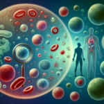
Imagine peering into the intricate corridors of the human mind—connecting neurons, pulsating synapses, and the electrical signals that orchestrate our thoughts and actions. The brain, with its approximately 86 billion neurons, is often described as one of the most complex structures in the known universe. When it comes to understanding neurological disorders, we must transcend traditional examination methods and illuminate the brain’s underpinnings using cutting-edge imaging techniques. This article explores the innovative imaging tools that allow us to scrutinize the brain under the microscope, unveiling critical insights into neurological disorders.
Understanding Neurological Disorders
Neurological disorders encompass a vast range of conditions impacting the nervous system, including Alzheimer’s disease, Parkinson’s disease, multiple sclerosis, and epilepsy. These disorders often present unique challenges, both in diagnosis and treatment, making advanced imaging an essential component of contemporary neuroscience.
The Rising Need for Innovative Imaging
Traditionally, neurological assessments relied heavily on clinical evaluations and neuropsychological tests, which can be subjective and limited. The need for more objective, illustrative tools became apparent as we sought to demystify the brain’s complexities. This led scientists and clinicians to innovate and adopt advanced imaging techniques that capture not just the structure of the brain but its intricate functionality.
The Evolution of Imaging Techniques
1. Magnetic Resonance Imaging (MRI)
Overview: MRI is a non-invasive imaging technique that offers a high-resolution view of the brain’s structure. Utilizing powerful magnets and radio waves, MRI allows for the visualization of soft tissue, making it invaluable for diagnosing and understanding disorders like tumors and strokes.
Case Study: Alzheimer’s Disease
A study published in Archives of General Psychiatry revealed that MRI scans could detect brain atrophy in patients diagnosed with early-stage Alzheimer’s disease, even before noticeable symptoms manifest. By monitoring hippocampal volume via MRI, researchers could better predict disease progression, emphasizing the technique’s pivotal role in early intervention strategies.
Table 1: MRI Advantages in Neurological Disorders
| Advantage | Description |
|---|---|
| High Resolution | Provides detailed images of brain structure |
| Non-invasive | Avoids the risks associated with surgical procedures |
| Functional Imaging | Can assess brain activity via functional MRI (fMRI) |
| Versatile Applications | Applicable to various neurological disorders |
2. Positron Emission Tomography (PET)
Overview: PET imaging functions through radioactive tracers that measure metabolic processes in the brain. This technique is particularly effective in highlighting areas of the brain that show abnormal activity related to various disorders.
Case Study: Parkinson’s Disease
A pivotal study in the Journal of Nuclear Medicine showed that PET imaging significantly enhanced the detection and differentiation between Parkinson’s disease and other parkinsonian syndromes, which was previously a clinical challenge. Using tracer uptake measurements, specialists could visualize dopamine transporter levels, leading to more accurate diagnoses.
3. Computational Tomography (CT)
Overview: CT imaging employs X-ray technology to create detailed images of the brain. While it is less effective for soft tissue than MRI, CT remains a critical tool in emergency scenarios, especially for detecting hemorrhages.
Case Study: Traumatic Brain Injury
In a study featured in Neurosurgery, researchers used CT scans to assess 2,000 patients with traumatic brain injury, illustrating how rapid CT evaluations could guide immediate treatment decisions, showcasing the method’s relevance in urgent care contexts.
4. Diffusion Tensor Imaging (DTI)
Overview: A specialized form of MRI, DTI maps out the white matter tracts of the brain, providing insight into the brain’s connectivity and the impact of disorders on neuronal pathways.
Case Study: Multiple Sclerosis
A landmark study in Neurology demonstrated that DTI could reveal disrupted white matter integrity in patients with multiple sclerosis, correlating with clinical outcomes. This study highlights DTI’s potential for tracking disease progression and evaluating treatment efficacy.
5. Electroencephalography (EEG)
Overview: EEG is an essential tool for observing electrical activity in the brain, often employed to diagnose epilepsy and sleep disorders.
Case Study: Epilepsy Diagnosis
A comprehensive study in Frontiers in Neurology illustrated how EEG could accurately identify seizure focus in patients unresponsive to standard treatments. By mapping electrical activity, clinicians can develop personalized treatment plans, underscoring the technique’s clinical utility.
Next-Generation Imaging Techniques
While the aforementioned techniques have revolutionized the neurological field, emerging technologies show promise for even deeper insights.
1. Functional Magnetic Resonance Imaging (fMRI)
Overview: fMRI measures brain activity by detecting changes associated with blood flow, providing a window into functioning networks.
Real-World Insight: Researchers conducted a study that mapped brain activity in patients during cognitive tasks, revealing the neural correlates of decision-making. Such insights could shape therapeutic approaches for cognitive impairments.
2. Optical Imaging
Overview: Confocal microscopy and two-photon microscopy allow for real-time, in-depth imaging of neural circuits at the cellular level, providing unmatched precision in analyzing neurobiology.
Case Study: Understanding Synaptic Changes
In recent research published in Nature Neuroscience, scientists utilized two-photon microscopy to observe synaptic changes in rodents associated with long-term memory formation. Such insights could illuminate mechanisms underlying memory disorders.
3. Machine Learning and Imaging
Overview: Integrating artificial intelligence with imaging technologies is paving the way for enhanced diagnostic accuracy. Machine learning algorithms can analyze vast datasets to identify patterns often imperceptible to human observers.
Case Study: Alzheimer’s Prediction
A recent study showcased how machine learning algorithms trained on MRI data could predict Alzheimer’s disease progression with remarkable accuracy, potentially revolutionizing early detection practices.
Challenges in Imaging Techniques
Despite the advantages of these cutting-edge tools, several challenges remain.
Accessibility and Cost
High-resolution imaging technologies often come with significant costs, limiting access for some populations. Ensuring equal access is essential to capitalize on these advancements.
Interpretation of Results
The complexity of neurological data requires specialists who can accurately interpret the findings. Training and education for healthcare professionals in advanced imaging interpretation are crucial.
Conclusion
The exploration of the brain under the microscope continues to evolve, revealing insights that were once unimaginable. By employing cutting-edge imaging techniques, we pave the way for better understanding, diagnosis, and management of neurological disorders. The future of neuroscience holds promise, with ongoing research poised to enhance our capabilities further.
As we stand at the intersection of technology and medicine, it’s a pivotal time to embrace these advancements. Whether it’s through technology infusion in clinical settings or nurturing a collaborative research environment, every step taken towards improving imaging techniques brings us closer to effectively addressing neurological disorders.
FAQs
1. What are the most common imaging techniques used in neurology?
Common techniques include MRI, CT, PET, DTI, and EEG, each serving distinct purposes in diagnosing and understanding neurological disorders.
2. How does MRI differ from CT imaging?
MRI uses magnetic fields and radio waves for imaging soft tissues, while CT uses X-rays, making MRI superior for examining the brain’s structure.
3. Can imaging techniques predict neurological disorders?
Techniques like fMRI and machine learning applications are showing promise in predicting disease progression by identifying patterns in brain activity.
4. Are these imaging techniques safe?
Most imaging methods, like MRI and fMRI, are non-invasive and considered safe. However, CT scans involve exposure to radiation, so usage must be justified.
5. How can I ensure equitable access to these imaging technologies?
Advocacy for policy changes, funding for medical facilities, and education about these technologies can help improve access across diverse populations.
By delving into the intricacies of the human brain through advanced imaging modalities, we are not just looking for answers; we are paving the way for hope, healing, and a better future for individuals grappling with neurological disorders. The brain under the microscope is not merely a subject of study—it is the future of health.