Introduction
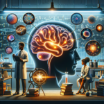
Imagine peering into the vast universe of the human mind, where complex thoughts, emotions, and memories swirl like stars in a galaxy. This once elusive capability is now closer to reality, thanks to the rapid advancements in imaging techniques that are redefining the field of neuroscience. Seeing Inside the Brain: How Imaging Techniques are Transforming Neuroscience offers us a glimpse into these revolutionary methods that not only enhance our understanding of the brain but also pave the way for breakthroughs in medical treatments and therapies.
As we embark on this journey, we’ll explore the various imaging techniques, their applications, and the transformative impact they hold for both research and clinical practices. From detecting neurological disorders to guiding surgical interventions, the ability to visualize brain activity opens a new chapter in our quest to understand the intricacies of the human mind.
The Landscape of Brain Imaging: An Overview
What is Brain Imaging?
Brain imaging refers to a set of techniques used to create visual representations of the interior of the brain. These methods provide researchers and clinicians with insights into brain structure, function, and metabolism. Seeing Inside the Brain: How Imaging Techniques are Transforming Neuroscience underscores the transition of these techniques from laboratory curiosities to essential tools in clinical settings.
Key Imaging Techniques
Magnetic Resonance Imaging (MRI)
- Overview: MRI uses strong magnetic fields and radio waves to generate images of organs and tissues inside the body. It is especially powerful for visualizing soft tissues.
- Application: Used to detect conditions such as tumors, brain injuries, and multiple sclerosis, emphasizing its role in clinical diagnosis.
Functional Magnetic Resonance Imaging (fMRI)
- Overview: A variant of MRI, fMRI measures brain activity by detecting changes in blood flow. It provides real-time data on brain function.
- Application: Utilized extensively in cognitive neuroscience to study brain networks involved in language, memory, and perception.
Technique Purpose Real-World Application MRI Structural Imaging Detecting tumors fMRI Functional Imaging Mapping brain activities during tasks Positron Emission Tomography (PET)
- Overview: PET scans use radioactive substances to visualize metabolic processes in the brain, providing insights into brain activity and chemistry.
- Application: Often employed in identifying Alzheimer’s disease and other neurodegenerative disorders.
- Electroencephalography (EEG)
- Overview: EEG captures electrical activity in the brain through electrodes placed on the scalp, offering a direct measurement of neuronal activity.
- Application: Crucial for diagnosing conditions like epilepsy and sleep disorders.
The Evolution of Imaging Techniques
Seeing Inside the Brain: How Imaging Techniques are Transforming Neuroscience reflects not only advancements in technology but also the shift in our understanding of the brain. Just a few decades ago, neurological research relied heavily on invasive methods. Imaging techniques have allowed us to peer inside the brain non-invasively, significantly enhancing both safety and comfort for patients.
Case Studies: Real-World Applications of Imaging Techniques
Case Study 1: Early Detection of Alzheimer’s Disease
A landmark study published in the journal Nature highlighted the role of PET scans in the early detection of Alzheimer’s disease. Researchers utilized PET imaging to observe amyloid-beta plaques in the brains of patients at various stages of cognitive decline. The findings revealed that individuals with subtle cognitive impairments exhibited amyloid deposits, allowing for early intervention strategies.
Relevance: This case underscores the potential of imaging techniques to transform diagnostics, enabling healthcare professionals to implement preventive measures before significant cognitive decline occurs.
Case Study 2: Mapping Language Processing
In a groundbreaking study by scientists at Harvard University, fMRI was employed to map the brain regions responsible for language processing. Participants engaged in tasks that required speaking, listening, and reading, while fMRI tracked blood flow to active brain areas. Results showed specific networks in the left hemisphere responsible for different language functions.
Relevance: This research illustrates how Seeing Inside the Brain: How Imaging Techniques are Transforming Neuroscience can enhance our understanding of complex cognitive processes and has implications for treating language disorders.
Case Study 3: Surgical Planning for Epilepsy
A clinical trial conducted by Stanford University illustrated how EEG and fMRI were combined to locate the source of seizures in patients with epilepsy. The integration of these imaging modalities allowed for precise mapping of epileptic foci, resulting in more targeted and successful surgical interventions.
Relevance: This case demonstrates how imaging techniques not only enhance diagnostic accuracy but also improve surgical outcomes, marking a significant advance in epilepsy treatment.
The Impact of Neuroimaging on Mental Health
Understanding Mental Disorders
The exploration of mental health issues such as depression and anxiety has been significantly enhanced by neuroimaging. Studies have employed both fMRI and PET to identify altered brain circuitry linked to these conditions. For instance, abnormalities in the prefrontal cortex and amygdala have been linked to heightened anxiety and mood disorders.
Innovative Treatments
Advancements in brain imaging are also guiding innovative treatment methods, including:
Transcranial Magnetic Stimulation (TMS): TMS utilizes magnetic fields to stimulate nerve cells in the brain. Neuroimaging helps in selecting the appropriate brain areas for treatment.
- Neurofeedback: This technique uses real-time fMRI data to help patients gain control over their mental states. By providing feedback on brain activity, patients can learn to alter patterns associated with anxiety and depression.
Challenges and Considerations in Brain Imaging
While the benefits of neuroimaging are vast, several challenges exist:
Cost and Accessibility
High-end imaging techniques like MRI and PET scans can be expensive and less accessible in rural or underserved areas. Efforts to make these technologies more affordable and widely available are crucial for broader impact.
Interpretation of Data
The complexity of brain imaging data requires skilled professionals for accurate interpretation. Misinterpretation can lead to incorrect diagnoses and treatments, underscoring the need for continued training for neurologists and radiologists.
Ethical Considerations
With the ability to visualize thoughts and emotions, ethical dilemmas arise regarding privacy and consent. Discussions around how to responsibly manage and use brain imaging data are essential as technology continues to advance.
Conclusion
Seeing Inside the Brain: How Imaging Techniques are Transforming Neuroscience is not just a technological evolution; it is a paradigm shift in understanding the human experience. The insights derived from imaging techniques have opened pathways to innovative treatments, enhanced diagnostic capabilities, and a more nuanced appreciation of the complexities of the brain.
As we continue to peel back the layers of the mind, it is imperative that we embrace these advancements while also addressing the challenges they pose. The future of neuroscience is bright, but it requires a commitment to ethical practices, accessibility, and ongoing education in the field.
Actionable Takeaways
- Stay Informed: Follow emerging research on neuroimaging and mental health treatments.
- Advocate for Accessibility: Support initiatives that aim to make brain imaging more widely available.
- Foster Ethical Discussions: Engage in dialogues about the implications of neuroimaging in society.
FAQs
1. How do imaging techniques differ from each other?
Imaging techniques vary in their methodologies. MRI provides structural visualization, fMRI measures brain activity through blood flow, PET scans observe metabolic processes, and EEG captures electrical activity. Each technique has unique applications and benefits.
2. Are there risks associated with brain imaging?
While non-invasive methods like MRI are generally safe, risks may include exposure to radiation in the case of PET scans, allergic reactions to contrast dyes, or discomfort during the procedure. It’s essential to consult with a medical professional regarding any concerns.
3. Can brain imaging help with mental health treatments?
Yes, neuroimaging can guide treatment choices by identifying brain activity patterns associated with various mental health conditions. Techniques like TMS and neurofeedback leverage imaging data to enhance therapy effectiveness.
4. How accessible are brain imaging techniques?
The accessibility of brain imaging can vary significantly depending on geographic location and healthcare systems. Efforts are ongoing to develop more affordable technologies and broaden access for underserved populations.
5. What ethical concerns are associated with neuroimaging?
Ethical issues include privacy concerns regarding brain data, consent, and the potential for misuse of information. It’s important to address these challenges by establishing guidelines to protect individuals’ rights and data security.
By harnessing the power of imaging techniques, the field of neuroscience is evolving at an unprecedented pace, promising a brighter future filled with understanding, innovation, and healing.